
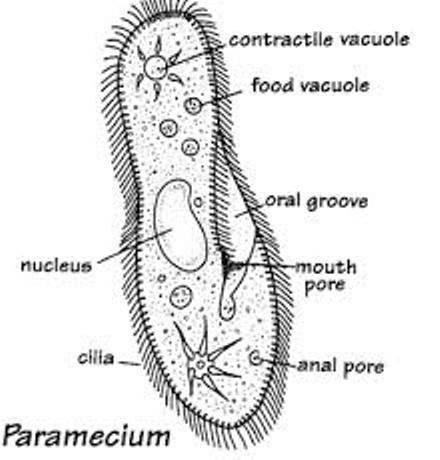
PARAMECIUM
Paramecium is widespread in freshwater, brackish, and marine environments and are often very abundant in stagnant basins and ponds. Because some species are readily cultivated and easily induced to conjugate and divide, it has been widely used in classrooms and laboratories to study biological processes.
Description of Paramecium
A paramecium is a unicellular (one cell) eukaryotic organism generally found in stagnant water. While very small, sometimes large paramecium can be seen as tiny specks darting around in a water sample. Paramecium can be about 0.5 mm long. Species of Paramecium range in size from 50 to 330 micrometres (0.0020 to 0.0130 in) in length. Cells are typically ovoid, elongate, foot- or cigar-shaped. The body of the cell is enclosed by a stiff but elastic membrane (pellicle), uniformly covered with simple cilia, hairlike organelles which act like tiny oars to move the organism in one direction. The pellicle gives the paramecium a definite shape but it is flexible enough to allow small shape changes. Nearly all species have closely spaced spindle-shaped trichocysts embedded deeply in the cellular envelope (cortex) that surrounds the organism. Typically, an anal pore (cytoproct) is located on the ventral surface, in the posterior half of the cell. In all species, there is a deep oral groove running from the anterior of the cell to its midpoint. This is lined with inconspicuous cilia which beat continuously, drawing food inside the cell. Paramecia live mainly by heterotrophy, feeding on bacteria and other small organisms.
Structure and Functions of Paramecium Parts
Pellicle – a membrane covering that protects the paramecium like skin
Cilia – hair like appendages that help the paramecium move food into the oral groove and also responsible for locomotion (movement)
Oral Groove – collects and directs food into the cell mouth also ingests nutrient.
Cell Mouth – opening for food
Anal Pore – disposes of waste
Contractile Vacuole – contracts and forces extra water out of the cell
Radiating Canals – paths to the contractile vacuole
Cytoplasm – intercellular fluid needed to contain vital cell parts
Trichocyst – used for defense
Gullet – forms food vacuoles
Food Vacuole – storage pocket for food. cavity of the paramecium responsible for digestion.
Macronucleus – larger nucleus which performs normal cell functions
Micronucleus – smaller nucleus which is responsible for cell division.
Paramecium Reproduction
Paramecium exhibit both sexual and asexual reproduction.
Asexual reproduction – This is the most common type of reproduction. The organism divides transversely. The macronucleus elongates and splits. Under ideal conditions, Paramecium can reproduce asexually two or three times a day.
Sexual Reproduction – Paramecium only reproduce sexually under stressful conditions. This occurs via gamete agglutination and fusion. Two Paramecium join together and their respective micronuclei undergo meiosis. Three of the resulting nuceli disintegrate, the fourth undergoes mitosis. Daughter nuclei fuse and the cells separate. The old macronucleus disintegrates and a new one is formed. This process is usually followed by asexual reproduction.
Paramecium Movement
The paramecium swims by beating the cilia. The paramecium moves by spiraling through the water on an invisible axis. For the paramecium to move backward, the cilia simply beat forward on an angle. If the paramecium runs into a solid object the cilia change direction and beat forward, causing the paramecium to go backward. The paramecium turns slightly and goes forward again. If it runs into the solid object again it will repeat this process until it can get past the object.
Paramecium Diet
Paramecium feed on microorganisms like bacteria, algae, and yeasts. The paramecium uses its cilia to sweep the food along with some water into the cell mouth after it falls into the oral groove. The food goes through the cell mouth into the gullet. When there is enough food in it so that it has reached a certain size it breaks away and forms a food vacuole. The food vacuole travels through the cell, through the back end first. As it moves along enzymes from the cytoplasm enter the vacuole and digest it. The digested food then goes into the cytoplasm and the vacuole gets smaller and smaller. When the vacuole reaches the anal pore the remaining undigested waste is removed. Paramecium may eject trichocyts when they detect food, in order to better capture their prey. These trichocyts are filled with protiens. Trichocysts can also be used as a method of self-defense. Paramecium are heterotrophs. Their common form of prey is bacteria. A single organism has the ability to eat 5,000 bacteria a day. They are also known to feed on yeasts, algae, and small protozoa. Paramecium capture their prey through phagocytosis
AMOEBA
The amoeba is a tiny, one-celled organism. You need a microscope to see most amoebas – the largest are only about 1 mm across. Amoebas live in fresh water (like puddle and ponds), in salt water, in wet soil, and in animals (including people). There are many different types of amoebas. The name amoeba comes from the Greek word amoibe, which means change. (Amoeba is sometimes spelled ameba.)
Description of Amoeba
This organism doesn’t have a rigid shape, but it is made of a flexible material that changes shape as needed. An amoeba is made of protoplasm, a viscous, clear material with a cell membrane separating the ectoplasm and the endoplasm, or the outer and inner parts of the cell. The endoplasm contains the nucleus of the cell.
An amoeba consists of a single blobby cell surrounded by a porous cell membrane. The amoeba “breathes” using this membrane – oxygen gas from the water passes in to the amoeba through the cell membrane and carbon dioxide gas leaves through it. A complex, jelly-like series of folded membranes called cytoplasm fills most of the cell. A large, disk-shaped nucleus within the amoeba controls the growth and reproduction of the amoeba.

Structure and Functions of Amoeba Parts
Cytoplasm – The cytoplasm is differentiated into Ectoplasm and endoplasm. The ectoplasm forms the outer and relatively firm layer lying just beneath the plasma lemma. It is a thin, clear (non-granular) and hyaline layer It is thickened into a hyaline cap at the advancing end at the tips of pseudopodia.
Plasma-lemma – Plasma-lemma is a very thin, delicate and elastic cell membrane of amoeba. It is composed of a double layer of lipid and protein molecules. This membrane is selectively permeable and regulates exchange of water, oxygen and carbon dioxide between the animal and the surrounding medium.
Cell membrane – the thin layer of protein and fat that surrounds the amoeba; it allows some substances to pass into the cell, and blocks other substances.
Contractile vacuole – The outer part of the endoplasm near the posterior end contains a clear, rounded and pulsating vacuole filled with a watery fluid. It is a cavity within the amoeba that excretes excess water and waste; the waste is brought to the cell membrane and is then eliminated from the amoeba.
Food vacuole – a cavity within the amoeba in which food is digested (broken down in order to be absorbed by the amoeba).
Nucleus – The nucleus has a firm nuclear membrane or nuclear envelope and contains a clear achromatic substance with minute chromatin granules or chromidia distributed uniformly near the surface. Nucleus is the major organelle of the amoeba, located centrally; it controls reproduction (it contains the chromosomes) and many other important functions (including eating and growth).
Pseudopodia – temporary “feet” that the amoeba uses to move around and to engulf food.
Amoeba Diet
An amoeba uses its pseudopodia to stretch out and reach food while surrounding it and pulling it back into the rest of the amoeba. The main components of an amoeba’s diet are bacteria and algae. The pseudopodia is one of the most important aspects to an amoeba. It helps the amoeba move, feed itself and reach anything that it needs to reach. Pseudopodia literally means fake foot, and this foot does nearly everything that the amoeba needs to do. It is the lifeblood of the amoeba. Pseudopodia are temporary finger like projections with blunt rounded tips which are constantly being given out or withdrawn by the body.
Amoeba Reproduction
Reproduction in amoeba chiefly occurs by asexual method, i.e., by binary fission, multiple fission and sporulation.
Binary Fission – In this process, the whole body divides into two daughter amoebae by mitosis. The division involves nuclear division (karyokinesis) followed by division of cytoplasm (cytokinesis). Division takes place under favourable conditions
Sporulation – Under un-favourable conditions amoeba reproduces by formation of spores internally. It starts with the breakdown of nuclear membrane and release of chromatin blocks into the cytoplasm. Each chromatin blocks acquires a nuclear membrane and becomes a small daughter nuclei. The newly formed nuclei get surrounded by cytoplasm to form amoebulae.
Multiple Fission – Under un-favourable conditions, amoeba divides by multiple fission. It withdraws its pseudopodia, becomes spherical and secretes three layered cyst around itself. Its nucleus undergoes repeated mitosis division forming 500- 600 daughter nuclei.
For more questions and explanations to your classwork, send an email to info@passnownow.com
1 thought on “Biology – Diagram of Paramecium and Amoeba with their Functions”
Awsmmm